Nidek MP-3 Microperimeter
Features
- Wide measurement range
- Auto tracking and auto alignment
- Feedback exam for visual rehabilitation
- Fixation test
- High resolution non-mydriatic fundus camera
- Gray scale
- Scotopic microperimetry (factory-installed option)
Wide measurement range
The MP-3 has a wider range of stimulus intensity, from 0 to 34 dB, compared to the MP-1. The MP-3 measures perimetric threshold values, even for normal eyes. A maximum stimulus luminance of 10,000 asb* allows evaluation of low-sensitivity.
*In accordance with ISO12866 measurement methods

Auto tracking and auto alignment
Auto tracking and auto alignment functions provide more accurate measurements increasing patient and operator comfort and efficiency. These functions allow easy follow-up and reduce variations between examiners, resulting in well-aligned follow up exams.
![]()
Feedback exam for visual rehabilitation
The visual rehabilitation mode trains low-vision patients who have lost foveal fixation to relocate their preferred retinal locus (PRL) to a different region, called the trained retinal locus (TRL). The TRL is predetermined by a physician, and fixation rehabilitation allows the patient better functional vision (i.e. reading speed) due to increased fixation stability and visual outcomes. Active flickering pattern stimulation and cheery music create an effective and pleasant training experience for the patient.

Image courtesy of the National Centre of Services and Research for the Prevention of Blindness and Rehabilitation of Visually Impaired - IAPB Italia Onlus, Rome - Italy
Fixation test
The MP-3 can measure fixation and determine the preferred retinal locus, simply by having the patient fixate on a target. Any change in fixation can be compared pre- and post-treatment because the patient’s eye is constantly tracked during microperimetry. This test allows evaluation of fixation in patients with central visual field defects and determines whether fixation improved after treatment.

High resolution non-mydriatic fundus camera
The 12-megapixel fundus camera in the MP-3 acquires high resolution images of retinal pathology and allows easy image acquisition.

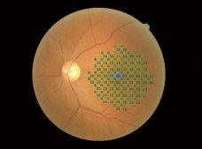
Gray scale
Visual sensitivity maps can be displayed in gray scale, similar to the conventional visual field reports. This display allows for an intuitive assessment of retinal sensitivity.
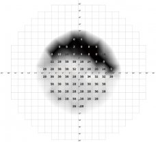
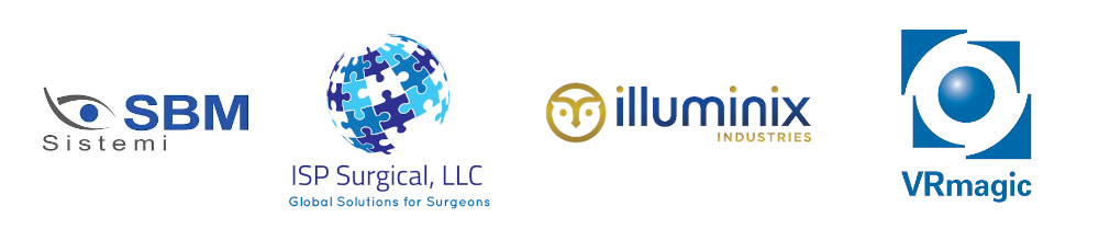
MP-3 images of pre- and post-treatment comparison
Case of anti-VEGF treatment for age-related macular degeneration (AMD)
Pre-treatment

- Circle at 2° Percentage of fixation points 66.1%
- Circle at 4° Percentage of fixation points 92.1%
- Mean sensitivity: 20.4
Post-treatment
- Circle at 2° Percentage of fixation points 68.1%
- Circle at 4° Percentage of fixation points 95.5%
- Mean sensitivity: 20.9
Scotopic microperimetry (factory-installed option)
Scotopic microperimetry is used to assess the changes in rod sensitivity of degenerative retinal diseases including age-related macular degeneration and some forms of retinitis pigmentosa. The type S provides scotopic microperimetry in addition to the standard functions of the MP-3.

Multimodal imaging
Various OCT modalities captured by the Mirante or RS-3000 Advance 2 can be registered with microperimetry.
OCT Angiography+Microperimetry

Normative DB+Microperimetry

Thickness Map+Microperimetry


Proud Distributors of