Nidek Angioscan Software
Features
- Comprehensive OCT-Angiography imaging and diagnostics
- The tracing HD plus function for accurate image capture
- Selectable definition and fine mode
- Wide area scan
- Vessel density map and perfusion density map
- Autodetection of FAZ and shape analysis
Comprehensive OCT-Angiography imaging and diagnostics
The simple interface provides seven slabs for the macula map / four slabs for the disc map with intuitive functionality and removal of projection artifacts. Segmentation into multiple slabs allows enhanced assessment of retinal microvasculature at specific depths and regions of interest.
Macula AngioScan: Macula Map -Vitreous, Superficial, Deep, Outer retina, ORCC, Choriocapillaris, Choroid
Optic Nerve Head AngioScan: Disc Map -Vitreous, Nerve head, Radial peripapillary capillary plexus (RPCP), Lamina cribrosa

The tracing HD plus function for accurate image capture
The tracing HD plus function tracks eye movement to maintain the same scan location on the SLO image for accurate image capture. Based on the clinical requirement, the tracing function can be set for high definition and high contrast imaging. Images can also be captured within seconds without the tracing function. Selectable definition and fine mode
Wide area scan
Up to 12 x 12 mm image can be captured.
Vessel density map and perfusion density map
Quantification of vessels in each layer provides metrics to assess disease progression and the effects of treatment. Quantitative analysis can be performed with vessel density map and perfusion density map.


Images courtesy of Chiba University Hospital Tohoku University
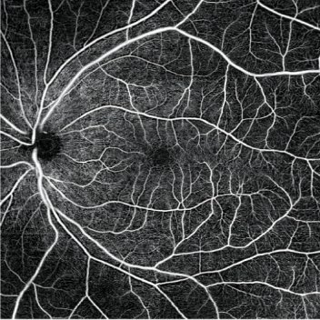
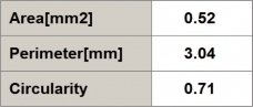
Autodetection of FAZ and shape analysis
Foveal Avascular Zone (FAZ) is automatically detected and shape metrics are provided for rapid assessment.


Proud Distributors of